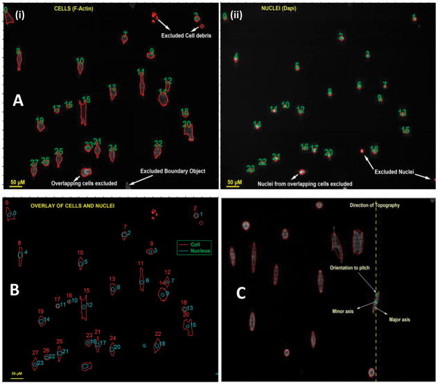
Figure 1.
(A) Figure illustrates the representative raw data from individual channels images of cells (actin) and nuclei (dapi), grown in EpiLife medium on 400 nm topography, with their outline identified. (B) Representative image overlay of the outline of cells and their corresponding nucleus. Figure demonstrates accurate matching of a cell to its nucleus. (C) Defining the axes of an object. Figure illustrates a representative of an ellipse defined around a cell to determine its major and minor axes, and hence its orientation to the underlying topography.

