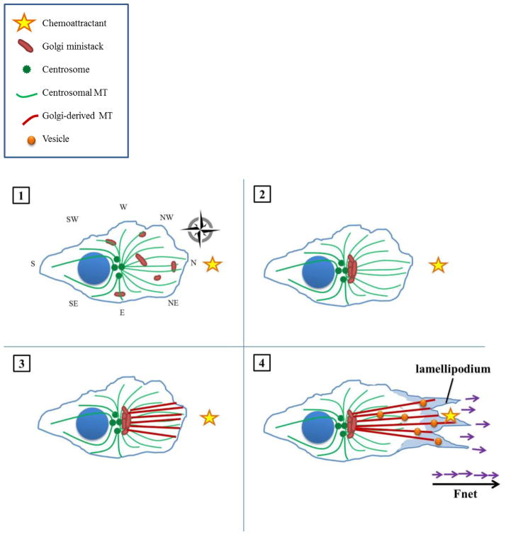
Figure 2. Enhanced directional cell migration in the presence of amplified centrosomes.
(1) In the interphase, pre-migratory cell exhibiting CA, Golgi ministacks are likewise scattered throughout the cytosol. The supernumerary centrosomes nucleate a similarly cell-wide, radially-symmetric array of microtubules; however, microtubule density is increased owing to the increased number of MTOCs as well as enhanced microtubule-nucleating capacity of individual MTOCs. The resultant amplified microtubule array constitutes an enhanced cellular compass (illustrated here as having, for instance, additional SW, NW, SE, and NE ordinal directions). A chemoattractant is arbitrarily situated due North. (2) Golgi ministacks are collected into a more compact, polarized, continuous mass in between the centrosomes and leading edge, as the cell is better able to gauge the precise location of the stimulus. (3) The Golgi directs its microtubules more precisely towards the stimulus, resulting in a more focused array. (4) Vesicles with migration- and invasion-promoting factors travel along microtubule tracks to a more well-defined point due North, allowing more forces (purple arrows) to be directed towards the stimulus and finally sum to a greater net force (bold black arrow). Thus, the cell can accelerate towards its stimulus more expeditiously.

