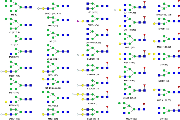
Fig. 3.
Symbol structures of the identified N-glycans. Numbering is in accordance with the peak numbering for the N-glycans in the fluorescence chromatograms and in Table 1. For structures with multiple peak assignments only one possible isomer is drawn. Symbols: inverted red triangles, l-fucose; blue squares, N-acetyl-d-glucosamine; green circles, d-mannose; yellow circles, d-galactose; blue diamonds, N-glycoylneuraminic acid; purple diamonds, N-acetyl-neuraminic acid

