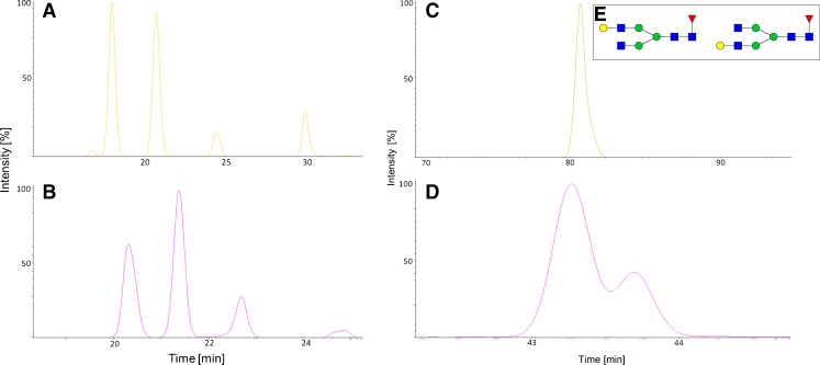
Fig. 7.
EICs of four structural M7 isomers (A, B) and G1F isomers (C, D) from mAb2. (A) EIC of 2-AB-labeled N-glycans. (B) EIC for the 2-AA-labeled glycans. Although selectivity is identical for the M7 isomers in both approaches, comparison of the peak areas reveals the order of elution changed for peaks 3 and 4. Selectivity is different for the G1F isomers. The 2-AB-labeled G1F elutes as one peak (C) whereas the 2-AA-labeled glycans (D) are separated into the two isomers. The terminal galactose residue (E) can be linked either to the α1,6 or the α1,3 branch of the bi-antennary N-glycan

