
Fig. 8.
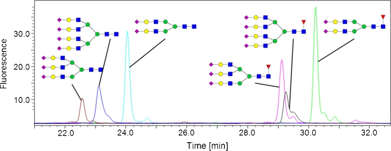
Overlay of fluorescence chromatograms derived from six different 2-AA-labeled sialic N-glycan standards. The appropriate structures are depicted. The grouping into non-fucosylated (three peaks on the left) and fucosylated (three peaks on the right) glycans is obvious