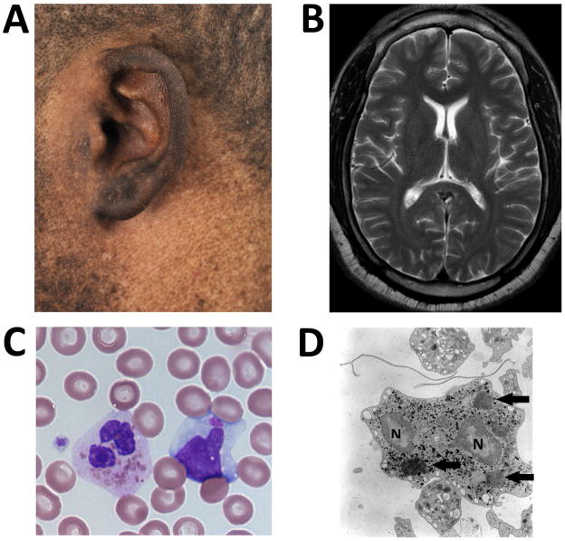
Figure 1.
A: Pigment dilution in this patient of African-American descent over the skin surrounding his left ear.
B: T2 weighted MRI image at the level of the head of the caudate nuclei.
C: Several pathognomonic giant intracellular inclusions within a neutrophil and a single giant inclusion in a lymphocyte on light microscopy of a peripheral blood smear.
D: Giant inclusions within the neutrophil on electron microscopy. Arrows identify giant inclusions. “N” identify portions of the segmented nucleus.

