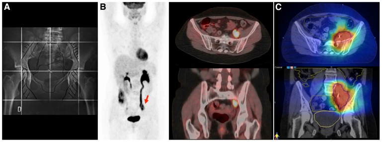
FIGURE 3.
A 43 year-old women with cervical cancer, FIGO IIB, without Kindly specify the same. evidence of macroscopic positive nodes at diagnosis. She was treated with chemotherapy and 3D conformal radiotherapy (45 Gy/1.8 Gy/fraction) followed by brachytherapy. (A) The initial radiotherapy field does not include the irradiation of common iliac nodes. (B) An FDG-PET/CT performed 2 years after primary treatment shows an isolated left iliac recurrence (arrow). This recurrence is observed near the border of the radiation field, which in the context of centrally controlled cervical cancer makes us suspect a component of marginal recurrence that typically arise immediately adjacent to the radiotherapy border. Surgical intervention was considered unfeasible and she underwent salvaged chemotherapy (carboplatin and taxane), followed by re-irradiation. (C) Re-irradiation was performed with helical tomotherapy using a hypofractionated schema of 15 daily fractions of 3.5 Gy. All tomotherapy plans were processed on VelocityAI to evaluate cumulative dose to normal tissue and organs at risk (OAR). Megavoltage computed tomography (MVCT) was performed every day before treatment to correct patient setup. The patient is alive without evidence of disease at the 3-year follow-up.

