Fig. 4.
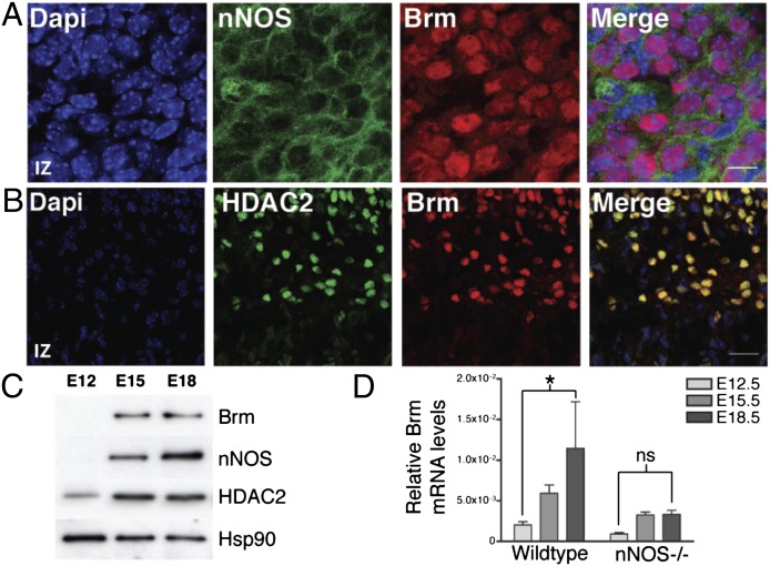
Expression of Brm is regulated during cortical development. (A and B) Images of sagittal sections of E18.5 cortex stained with DAPI (blue), anti-Brm (red), anti-nNOS (green; A), or anti-HDAC2 (green; B). (Scale bars, 10 µm in A and 30 µm in B.) n = 3. (C) Western blot analysis of Brm, nNOS, HDAC2, and Hsp90 in cortical lysates from E12.5, E15.5, and E18.5 embryos. n = 3. (D) qRT-PCR analysis of Brm mRNA levels normalized to Rpl11. mRNA was isolated from cortices of either wild-type or nNOS−/− embryos at the indicated stages. Averages are SEM; *P < 0.05; ns, nonsignificant; two-way ANOVA, Bonferroni post hoc test; n = 4.

