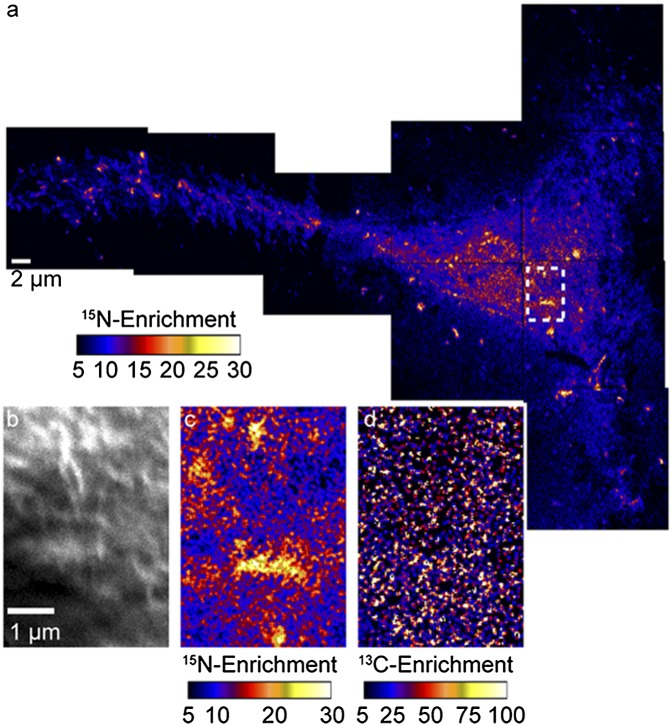
Fig. P1.
NanoSIMS images of the surface of a Clone 15 fibroblast cell. (A) The 15N-enrichment image of the entire dorsal surface of the cell shows 15N-sphingolipid-rich domains within the plasma membrane, evidenced by statistically significant local elevations in the 15N-enrichment. (B) The secondary electron image that corresponds to the region outlined in A shows the cell has normal morphology. (C) The 13C-enrichment image of the same region confirms the membrane was intact and the sample preparation and NanoSIMS imaging did not create artifactual lipid clustering. (D) Domains enriched with 15N-sphingolipids are visible in the enlarged 15N-enrichment image of the region outlined in A.

