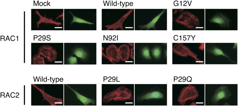
Fig. 2.
Actin reorganization induced by the RAC1/RAC2 mutants. 3T3 cells infected with retroviruses encoding enhanced green fluorescent protein (EGFP) as well as wild-type or mutant forms of RAC1 or RAC2 were stained with Alexa Fluor 594-labeled phalloidin to visualize actin organization (Left image of each pair). The same cells were also examined for EGFP fluorescence (Right image of each pair). (Scale bars, 20 µm.)

