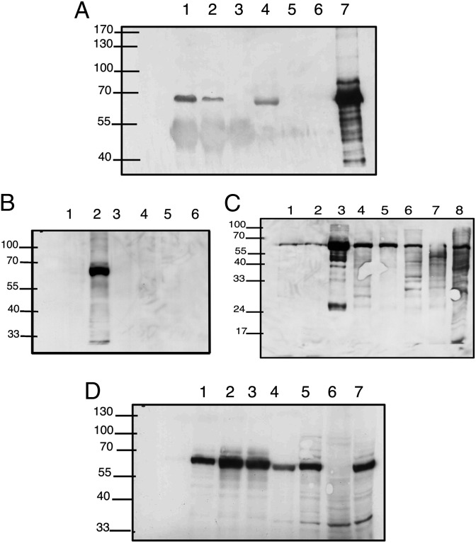
Fig. 5.
(A) Immunoprecipitation of 67-kDa band with anti-human serum albumin antibody. Nuclear (lane 1) or cytoplasmic (lane 2) lysates of human CD4 T cells treated with penicillin (control: no lysate, lane 3) were incubated with rabbit polyclonal anti-human albumin (Sigma), precipitated with protein A Sepharose, and run on an SDS gel. Lysates without the protein A bound complex (lane 4, nuclear; lane 5, cytoplasmic; lane 6, no lysate control; lane 7, positive control) were run. Western blot analysis was performed with monoclonal anti-penicillin (Pen 9). IP of the cytoplasmic fraction with anti-human albumin resulted in the disappearance of the penicilloyl band, indicating that the only labeled protein was albumin. (B) T cells express albumin modifiable by penicillin. Human CD3 cells were stimulated in vitro with PMA and ionomycin in the presence of the indicated antibiotics for 48 h. The cells were collected, washed, and lysed in lysis buffer, and the lysates were separated in SDS/PAGE. Proteins were transferred to nitrocellulose and probed with Pen 9 in standard Western blots. The Pen 9-labeled 67-kDa band is present only in penicillin-treated T cells. Lane 1 cells, untreated with antibiotics; lane 2, penicillin; lane 3, ampicillin; lane 4, cefuroxime; lane 5, chloramphenicol; and lane 6, vancomycin. (C) Detection of in vivo penicillin-labeled proteins. Lewis rats were injected with penicillin G; 120 min after injection, the rats were killed and various tissues were subjected to SDS/PAGE and Western blotting with monoclonal Pen 9 antibody. The 67-kDa band is present in all tissues and is most abundant in the serum sample. Lane 1, thymus; lane 2, spleen; lane 3, serum; lane 4, kidney; lane 5, intestine; lane 6, liver; lane 7, lung; lane 8, positive control human PBL. (D) Western blot analysis of various cell lines treated with penicillin shows the dominant 67-kDa band in most samples. Lane 1, mouse mesenchymal stem cells; lane 2, mouse immature dendritic cells; lane 3, mouse mature dendritic cells; lane 4, Jurkat; lane 5, human acute lymphoblastic leukemia (ALL) line MOLT-4; lane 6, FAO rat hepatoma cell line; lane 7, CEM human ALL line. The band is absent from a hepatoma cell line that was documented to have undergone dedifferentiation and lost its albumin expression (16).

