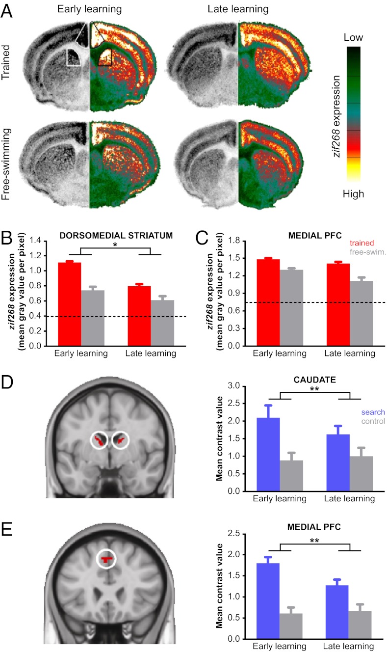
Fig. 2.
Dorsomedial striatum and medial prefrontal cortex support early place learning in human and mouse. (A) Coronal sections from mouse displaying zif268 expression during early and late learning in experimental and free-swimming control groups. The left hemisphere shows the original autoradiogram in gray scale and is matched on the right by its pseudocolor counterpart. The color scale bar ranges from no signal (0, dark green) to maximum signal (255, white). Striatal and prefrontal subdivisions in mouse were based on known anatomical connectivity (Fig. S4 A and B). (B) A larger reduction in zif268 expression between early and late learning was observed in the experimental groups than in the free-swimming controls, suggesting a specific role for this region during early learning. (C) zif268 expression in medial prefrontal cortex decreased from early to late in both experimental and free-swimming control groups. (D) Posterior dorsomedial striatum in human (image displayed at Montreal Neurological Institute coordinate y = −3) responded more strongly to search trials than control trials (P < 0.05, FWE corrected). Subdivisions within human striatum were based on prior knowledge regarding functional differences (Fig. S4 C and D). (E) Medial prefrontal cortex in human (y = 24) responded more strongly to search trials than control trials (P < 0.05, FWE corrected) and was functionally connected to the dorsomedial striatum at rest (P < 0.05, FDR corrected). Mean contrast values were extracted from the activations shown in the left of D and E and are plotted to the right of each image. Error bars represent SEM. *P < 0.05; **P < 0.01.

