
Figure 2.
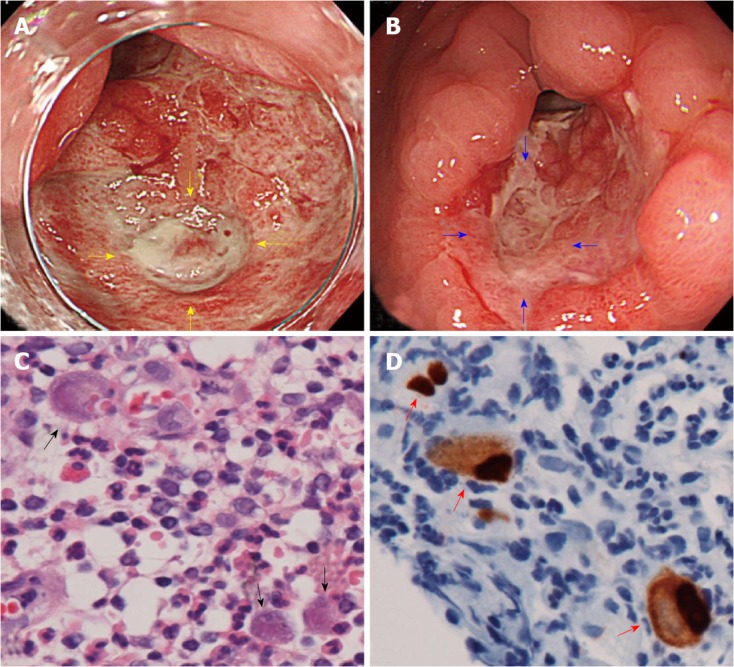
Endoscopic findings of cytomegalovirus associated ulcer and microscopic examination. A: Artificial ulcer on postoperative day 12, showing the formation of abundant granulation tissue and a 10-mm-deep ulcer at the center of the granulation tissue (yellow arrows); B: The healing process of the deep ulcer (blue arrows) on postoperative day 15; C: A biopsy from the deeper ulcer margin revealed large cells with intranuclear inclusion bodies (black arrow, HE staining, × 600); D: Large cells with intranuclear inclusion bodies stained positive for anti-cytomegalovirus (CMV) antibodies (red arrows, anti-CMV antibody immunohistochemical staining, × 600).