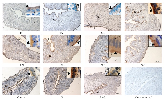
Figure 4.
Immunolocalization of NHE1 in steroid replaced ovariectomized rats and in rats at different phases of the oestrous cycle. NHE1 expression was the highest under P dominance predominantly at the apical membrane. Magnifications of 10X and 100X (in the upper right corner). 0.2E: 0.2 μg 17β-oestradiol, 2E: 2 μg 17β-oestradiol, 20E: 20 μg 17β-oestradiol, 50E: 50 μg 17β-oestradiol, P: progesterone, E + P: 0.2 μg 17β-oestradiol + progesterone. Ps: proestrus, Es: estrus, Ms: metestrus, Ds: diestrus. n = 6 rats per group. Incubation with a non-immune peptide was used as a negative control to check for the specificity of the antibodies. No staining was observed in this experiment.

