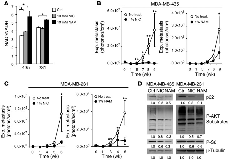
Figure 6. NAD+ precursor treatment inhibits metastatic activity.
(A) NAD+ precursor treatment enhanced the NAD+/NADH ratio in cultured MDA-MB-435 and MDA-MB-231 parental cells. NAD+/NADH levels were measured after 3 days of cell treatment with 10 mM NIC or NAM in complete medium. n = 3 independent experiments. (B and C) NAD+ precursor treatment of experimental mice inhibited lung metastasis. Lung colonization by MDA-MB-435 (B) or MDA-MB-231 (C) parental cells (2.5 × 10⁵ i.v. each) in mice treated with NIC or NAM (1% in the drinking water ad libitum throughout the experiment). Controls received no treatment (plain drinking water at same pH). Metastatic growth was measured by repeated noninvasive bioluminescence imaging. n = 6 per group. (D) NIC or NAM treatment influenced mTORC1 activity and autophagy. Western blot analysis for p62, phospho-AKT substrates, and phospho-S6Ser240/244 in MDA-MB-435 or MDA-MB-231 parental cells with or without 48 hours of treatment with 10 mM NIC or NAM. β-Tubulin served as protein loading control. Signal quantification, measured by infrared imaging (total of detectable bands) and expressed relative to control, is shown below. Results are representative of 3 independent experiments. *P < 0.05, **P < 0.01, unpaired 2-tailed Student’s t test (A) or nonparametric Mann-Whitney test (B and C).

