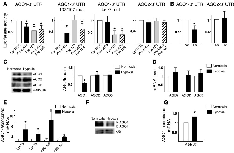
Figure 3. Posttranscriptional targeting of AGO1 mRNA in AGO1-mediated miRISC.
(A) HEK293 cells were transfected with the WT Luc-AGO1–3′ UTR (WT), Luc-AGO1–3′ UTR with miR-103/107 or Let-7 target sites mutated (mut), or Luc-AGO2–3′ UTR, together with pre–Let-7e (40 nM), pre-103 (40 nM), pre–Let-7e and pre-103 (20 nM each), or control RNA (40 nM). (B) Bovine aortic ECs (BAECs) transfected with Luc-AGO1–3′ UTR or -AGO2–3′ UTR were subjected to normoxia (Nx) or hypoxia (Hx). CMV–β-gal was cotransfected in all groups as a transfection control. Luciferase activity was normalized to that of β-gal. (C–G) HUVECs were subjected to normoxia or hypoxia. (C and D) Western blot and qPCR analyses of protein and mRNA levels of AGO1–3. (E–G) AGO1 was immunoprecipitated from cell lysates. The immunoprecipitates were subjected to AGO1 immunoblotting (F) and the AGO1-associated miRNAs and AGO1 mRNA were quantified by qPCR (E and G). Data represent mean ± SD from 3 independent experiments. *P < 0.05 compared with control RNA group in A or normoxia group in B–G.

