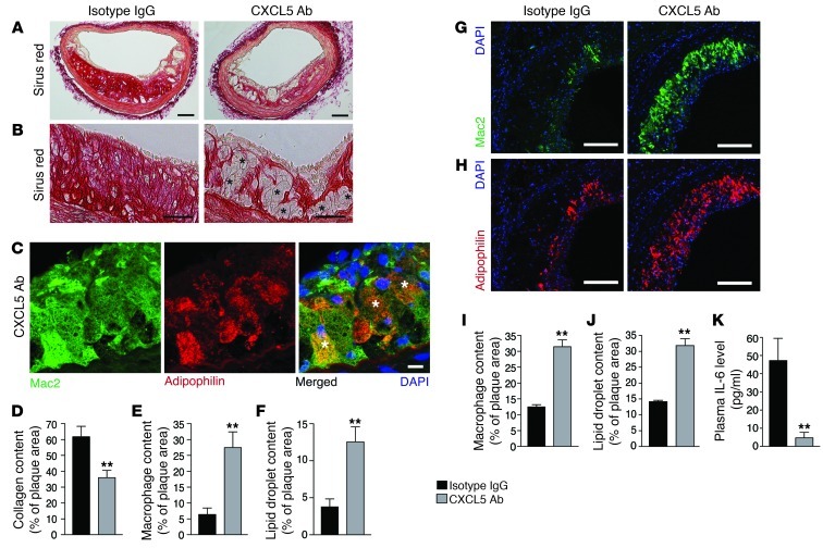
Figure 2. Blockade of CXCL5 is associated with macrophage foam cell accumulation in atherosclerotic plaques.
Apoe–/– mice were fed a WD and treated with either IgG isotype control (IgG) or anti-CXCL5 Ab (CXCL5 Ab) for 12 weeks. (A and B) Representative images from brachiocephalic artery lesions of picrosirius red staining for collagen detection. Black asterisks indicate some foam cells. (C) Representative images of macrophages (Mac2 immunostaining) containing lipid droplets (adipophilin immunostaining) from anti-CXCL5 Ab-treated brachiocephalic artery lesions. White asterisks indicate double-positive cells. (D–F) Quantification of (D) sirius red staining, (E) Mac2, and (F) adipophilin immunostaining in brachiocephalic artery lesions. Representative images from aortic root lesions of (G) Mac2 and (H) adipophilin immunostaining. Quantification of (I) Mac2 and (J) adipophilin immunostaining in aortic root lesions. (K) Quantification of plasma IL-6 by ELISA. Data in D–F and I–K represent mean ± SEM. n =7–8. **P < 0.01. Scale bars: 100 μm (A, G, and H); 50 μm (B); 10 μm (C).

