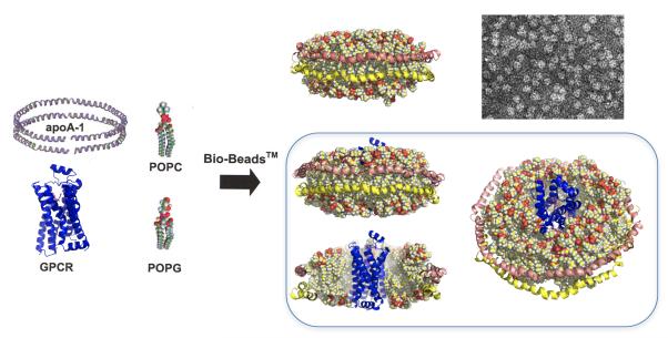
Figure 1. Reconstitution of a prototypical GPCR into rHDL particle.
Illustration of the procedure for the reconstitution of the b2AR (PDB: 2RH1) into rHDL particles. Detergent-solubilized purified lipids and purified β2AR are incubated with purified apolipoprotein A1 (apo-A1) as described in the text. Bilayer formation and self-assembly of the rHDL particle accompanies the detergent removal step through the addition of Biobeads™. Both empty and β2AR-containg rHDL particles are illustrated (the latter are illustrated from multiple perspectives). Also illustrated is the electron micrograph of a typical rHDL preparation (Whorton and Sunahara, unpublished). The coordinates for the rHDL particle were based on the model reported by Segrest et al (58) and used with the permission of Dr. Stephan Harvey (Georgia Institute of Technology).

