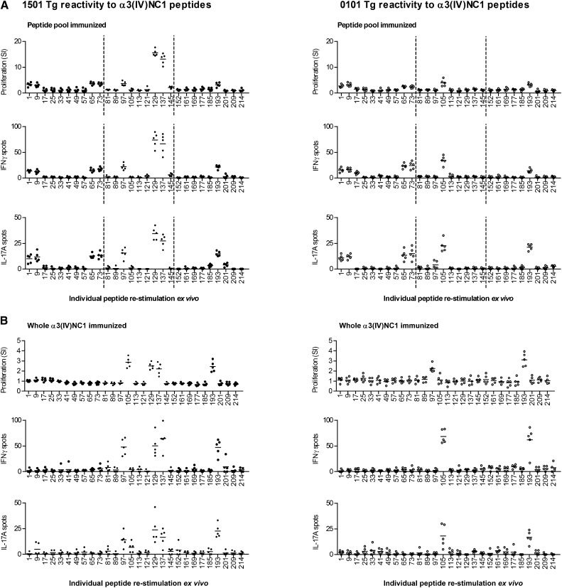
Figure 1.
Distribution of the α3(IV)NC1 T cell epitopes in HLA-DRB1*15:01 and HLA-DRB1*01:01 transgenic mice. Mice deficient in murine MHC II but possessing human DRB1*15:01 (1501 Tg) or DRB1*01:01 (0101 Tg) after immunization with either (A) peptide pools (with each pool encompassing a third of α3(IV)NC1, dotted lines) or (B) whole α3(IV)NC1. Reactive peptides are defined by re-stimulation of draining lymph node cells by ex vivo proliferation, IFN-γ ELISPOT, and IL-17A assays. Each dot represents the response from a mouse (n=5 per group). Numbers on the x axis represent the α3(IV)NC1 N-terminal number of a 20-mer. Several areas of reactivity are present, but one area of strong reactivity, α3129–148 and α3137–156 (corresponding to one of two epitopes found in humans with anti-GBM disease), is present only in 1501 Tg mice.

