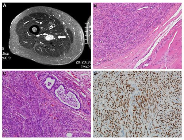
Figure 1.
Benign metastasizing leiomyoma. (A) MRI of well-circumscribed thigh mass (white arrow). (B) Thigh mass exhibiting bland, relatively cellular, smooth muscle proliferation without atypia, necrosis, or mitotic activity. (C) Similar histopathologic appearance for the pleuropulmonary lesion. (D) Strong, diffuse nuclear staining for estrogen receptor in the pleuropulmonary lesion.

