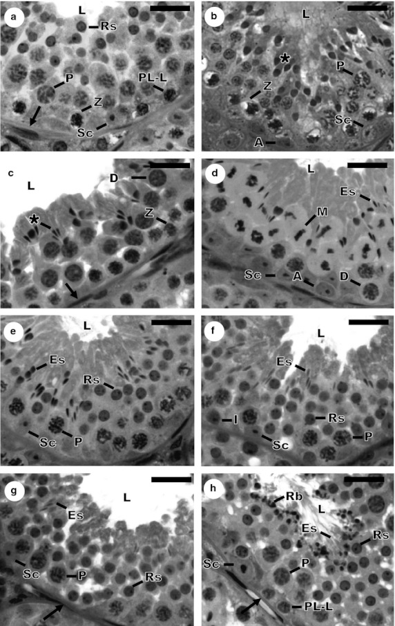
Fig. 1.

Histological cross-sections of seminiferous tubules showing the eight stages of the seminiferous epithelium cycle in Sturnira lilium, according to the tubular morphology method. (a) Stage 1; (b) Stage 2; (c) Stage 3; (d) Stage 4; (e) Stage 5; (f) Stage 6; (g) Stage 7; (h) Stage 8. Sc, Sertoli cell; A, type A spermatogonia; I, intermediate spermatogonia; PL-L, primary spermatocyte in Pre-leptotene to leptotene; Z, primary spermatocyte in zygotene; P, primary spermatocyte in pachytene; D, primary spermatocyte in diplotene; M, metaphase figure; Rs, round spermatid; *, elongating spermatid; Es, elongated spermatid; →, tunica propria; L, lumen of the seminiferous tubule; Rb, residual body. Scale bars: 10 μm.
