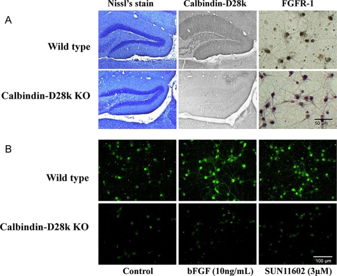
Figure 5.

Immunocytochemical characterization of brain neurons from wild-type (WT) and homozygous calbindin-knockout (Calb–/–) mice. (A) Immunoreactivity to calbindin-D28k and fibroblast growth factor receptor-1 (FGFR-1) in neurons from WT and Calb–/– mice. In order to show the gene deletion of calbindin-D28k, typical brain tissue sections were employed for confirmation. Primary cultures from the brains of WT and Calb–/– mice displayed that they contained neurons with immunoreactive FGFR-1. (B) Increased levels of expression of calbindin-D28k by bFGF and SUN11602 in primary cultures of cerebrocortical neurons from WT and Calb–/– mice. Culture neurons that were grown on coverslips in the presence of 10 ng/mL bFGF or 3 μM SUN11602 for 48 h were fixed and immunostained with a calbindin-D28k antibody. Calbindin D28k-immunoreactivity was mostly eliminated from the cultures of cerebrocortical neurons from Calb–/– mice.
