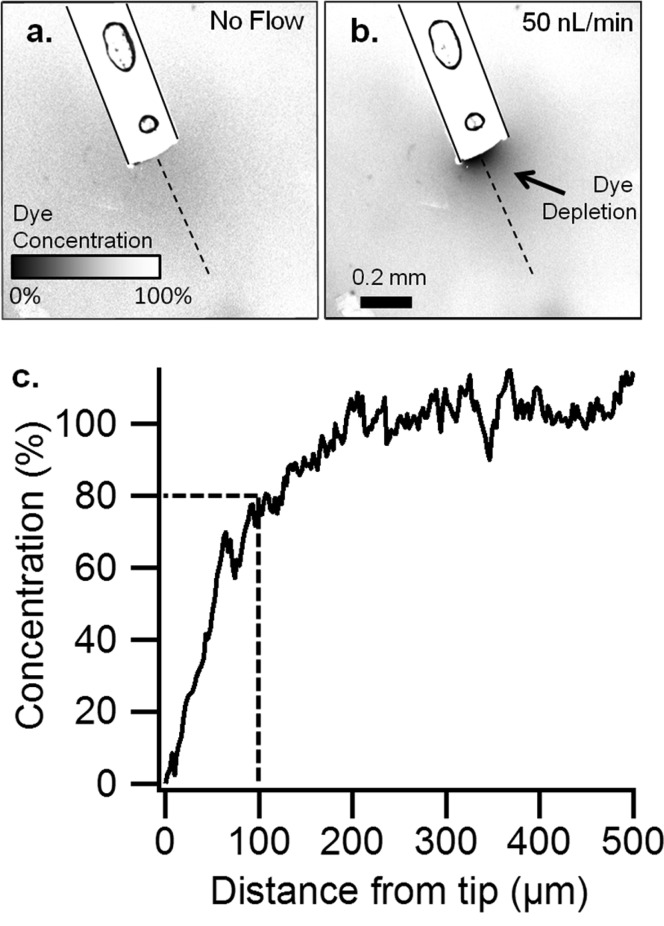
Figure 2.
Determination of spatial resolution by low flow push–pull perfusion in vitro. Confocal microscopy of a probe (outlined) in agar containing 1 μM resorufin (a) without sampling and (b) push–pull sampling at 50 nL/min. (Circles are air bubbles within the probe epoxy.) (c) The relative concentration of resorufin in a linear path (dashes in A,B) from the tip of the probe, measured by the difference in fluorescence. Scale bar is 200 μm.

