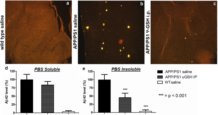
Figure 5.
Immunohistochemical detection of Aβ1–42 in ψ-GSH-treated mouse brain, magnification 10×: (a) wild-type (WT) saline control; (b) APP/PS1 saline; (c) APP/PS1 ψ-GSH ip. Amyloid plaques are observed as intensely yellow stained regions in sagital sections of brain. Mice treated with ψ-GSH showed dramatic reduction in size and number of plaques. (d, e) Quantitation of the effect of ψ-GSH treatment on amyloid load in APP/PS1 mice. Aβ1–42 levels in the brain homogenate of mice treated with saline or ψ-GSH for 12 weeks were measured by ELISA as described in Methods. (d) PBS-soluble Aβ1–42 levels were not different within treatments groups. (e) Significant reduction in PBS-insoluble (guanidine-soluble) Aβ1–42 was observed in ψ-GSH-treated APP/PS1 brain (∗∗∗, p < 0.0001 compared with saline-treated controls).

