Figure 2.
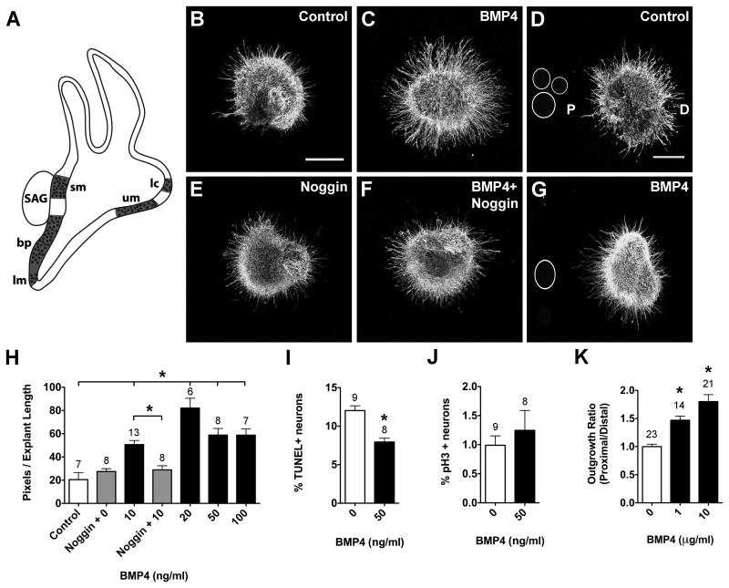
BMP4 promotes neuron survival and neurite outgrowth in HH20-25 SAGs cultured for 24 hours.
Summarized expression of Bmp4 transcripts (A) in the developing chick inner ear during stages of SAG neuron axon pathfinding and sensory organ innervation; based on (Wu and Oh, 1996). Prosensory regions are represented by dotted areas (here and in subsequent figures) and include the basilar papilla (bp), lateral crista (lc), lagena macula (lm), saccular macula (sm) and utricular macula (um). SAG explants cultured with vehicle (Control; B), 10 ng/ml BMP4 (C), 1 μg/ml Noggin (E), or 10 ng/ml BMP4 plus 1 μg/ml Noggin (E). SAG explants co-cultured with beads soaked in PBS (Control; D) or 10 μg/ml BMP4 (G). Scale bar = 200 μm. Quantification of the average pixel number for protein-treated SAG explants (H), the percentage of TUNEL positive SAG neurons (I) and the mitotic index of SAG neurons (J). Quantification of neurite outgrowth for SAG-bead co-cultures (K). In all figures, the following labeling conventions are used: bars represent the mean (±SE) computed from explants within a treatment group; sample numbers for each treatment group are above the bars; the outgrowth ratio is the average proximal/distal ratio of pixels per explant length for SAG-bead co-cultures with D as distal quadrant and P as proximal quadrant. In this figure, *p < 0.0001 significantly different from controls.

