Figure 7.
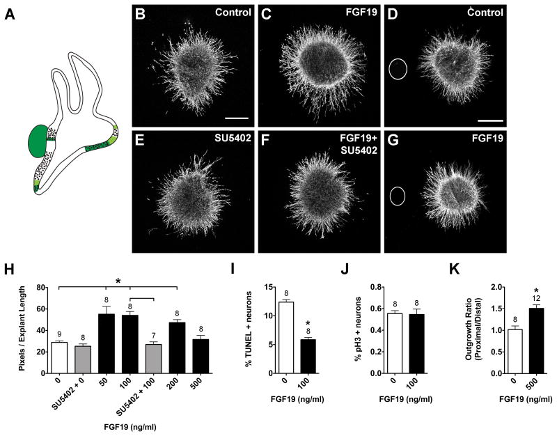
FGF19 promotes neuron survival and neurite outgrowth in HH20-25 SAGs cultured for 24 hours.
Summarized expression patterns of Fgf19 transcripts (A). Fgf19 is strongly expressed in the utricular macula, lagena macula, SAG, and displays weaker expression in some of the prosensory region borders, based on (Sanchez-Calderon et al., 2007). SAG explants cultured with vehicle (Control; B), 100 ng/ml FGF19 (C), 0.1 μM SU5402 (E), or 100 ng/ml FGF19 plus 0.1 μM SU5402 (F). SAG explants co-cultured with beads soaked in PBS (D) or 500 ng/ml FGF19 (G). Scale bar = 200 μm. Quantification of the average pixel number for SAG explants (H), the percentage of TUNEL positive neurons (I) and the mitotic index (J). Quantification of neurite outgrowth for SAG-bead co-cultures (K). See legend to figure 1 for labeling conventions. *p<0.0001 significantly different from controls.

