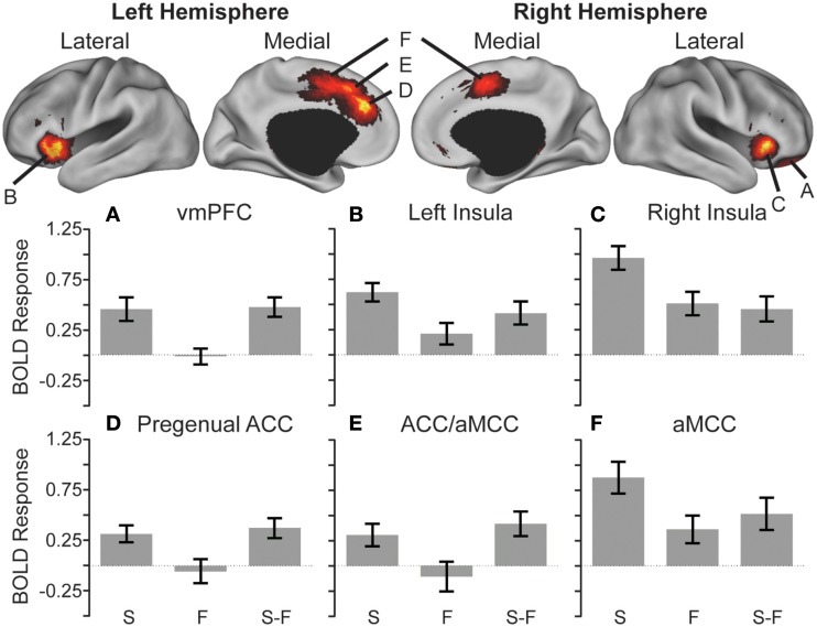
Figure 3.
Anticipatory activations showing the effect of stimulus. Cortical surface renderings of snake (S) minus fish (F) contrast using multi-fiducial mapping in CARET with the strongest voxel within 2.5 mm of the surface (Van Essen, 2005). Results were thresholded at p < 0.05 cluster corrected. Bottom: heightened anticipatory activity reflected in greater activation in the ventromedial prefrontal cortex (vmPFC) (A), bilateral anterior insula (B,C), pregenual anterior cingulate cortex (ACC) (D), regions spanning from the ACC to the anterior mid-cingulate cortex (aMCC) (E), and bilateral aMCC (F) preceding snake videos compared to fish videos (S > F).

