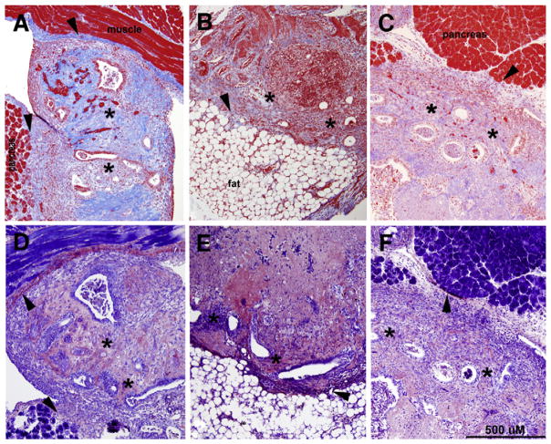
FIGURE 2.
Masson trichrome staining (A–C) or phosphotungstic acid hematoxylin staining (PTAH; D–F) of adhesions/lesions removed from control (A, D) or mice provided 10% (B, E) or 5% (E, F) fish oil–supplemented diet. Masson trichrome staining results in red-blue cell nuclei, red cytoplasm, and bright blue collagen. Notably, the intensity of the blue stain has been shown to correlate with the extent of collagen present (29). Within tissues obtained from fish oil–supplemented mice, the reduced amount and intensity of blue staining suggests that the amount of collagen is also reduced (B, C). PTAH staining of near sister sections was conducted to assess the presence of fibrin (which appears as a dark blue stain) and was similar in all groups (D–F). The asterisks denote areas containing endometrial glands and stroma, indicative of endometriotic-like lesions, and arrowheads mark sites of abnormal tissue attachment (adhesion). Photomicrographs are representative results of multiple tissues from at least four mice per group. Original magnification ×100.
Herington. Fish oil inhibition of adhesions. Fertil Steril 2013.

