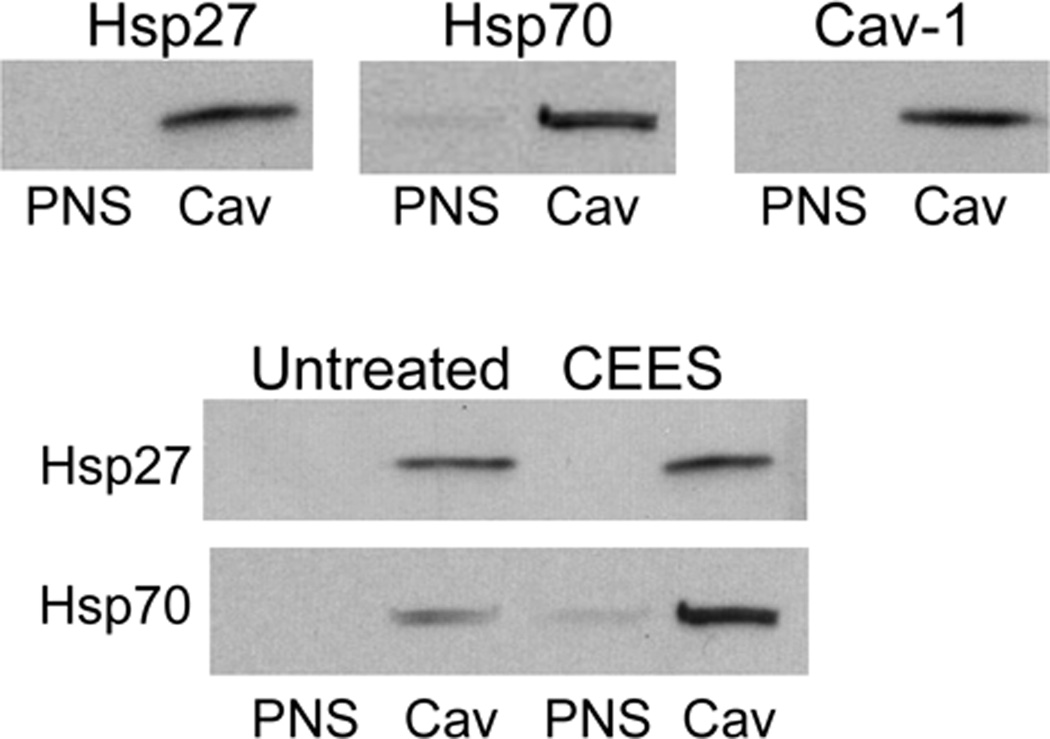
Figure 8. Localization of Hsp27 and Hsp70 in caveolae.
Mouse keratinocyte skin constructs were exposed to CEES (300 µM) or control. After 24 hr, caveolar (Cav) and post-nuclear supernatant (PNS) subcellular fractions were isolated from the cells using sucrose density gradient centrifugation as described in the Materials and Methods. Upper panel: Cav and PNS fractions were assayed for Hsp27 and Hsp70 by Western blotting. The relative purity of the caveolar fractions was determined by Western blot analysis using caveolin-1 antibodies. Lower panel: Effect of CEEES on Hsp27 and Hsp70 expression in cav and PNS subcellular fractions isolated from control and CEES-treated mouse keratinocytes.

