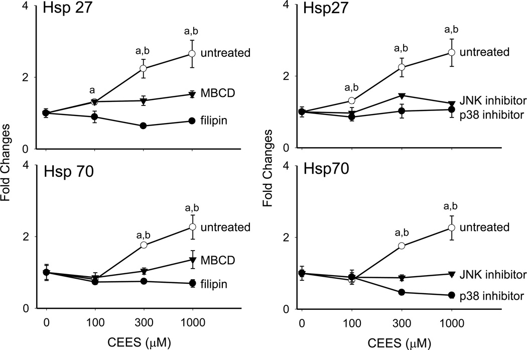
Figure 9. Effects of caveolae and MAP kinase inhibitors on CEES-induced expression of Hsp27 and Hsp70.
Mouse keratinocyte skin constructs were preincubated with the caveolae inhibitors, filipin (10 µM), and MBCD (5 mM), or control for 30 min, or JNK (SP600125, 20 µM) and p38 (SB203580, 10 µM) MAP kinase inhibitors or control for 3 hr, and then exposed to CEES (0, 100, 300 or 1000 µM). After 24 hr, mRNA was isolated from the cells and analyzed by real-time PCR. Data are presented as fold change in gene expression relative to untreated cells. Left panel: Effects of caveolae inhibition on Hsp27 and Hsp70 expression. aSignificantly (p < 0.05) different from control (filipin); bSignificantly different (p < 0.05) from control (MBCD). Right panel: Effects of MAP kinase inhibition on Hsp27 and Hsp70 expression. aSignificantly (p < 0.05) different from control (p38 inhibitor); bSignificantly different (p < 0.05) from control (JNK inhibitor).

