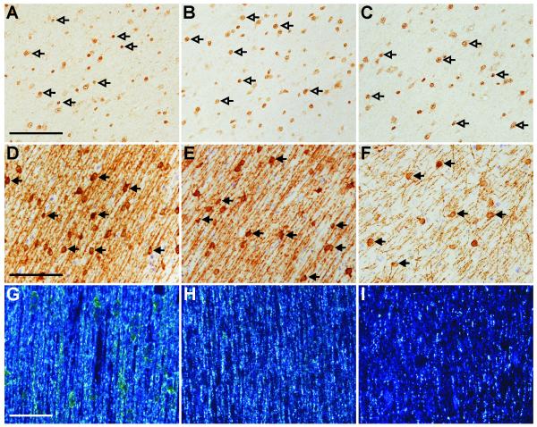
Figure 1. Effect of steroids on myelination.
Areal density of Olig2-IR oligodendrocytes (open arrows) did not differ between control (A), single course (B) and repeated course (C) groups, demonstrated in the subcortical white matter. The areal density of MBP-IR cells (closed arrows) in the white matter was reduced in the repeated course animals (F) compared to both the control animals (D) and the single course animals (E). Intensity of MBP-IR as viewed under darkfield illumination was reduced in the repeated course groups (I) compared to the control (G) and the single course (H) groups. Scale bars: A-C=100μm; D-F=100μm; G-I=50μm.

