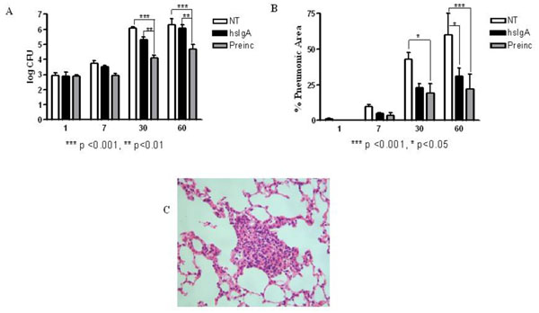
Figure 2.

Determination of bacterial load (A) and pneumonic area (B) in lungs of mice which were untreated (NT) and those treated with hsIgA (hsIgA), after challenge with M. tuberculosis H37Rv by intratracheal route 2 hrs after inoculation. Another group received M. tuberculosis preincubated with hsIgA (preinc). Granulomas of preincubated group 2 months after challenge with M. tuberculosis, visualized by hematoxilin-eosin staining (25x) (C). The morphometric study was carried out with light microscopy using Leica Q-win System Software. All data was analysed using GraphPad Prisma 4 Software. Each bar represents the mean of three samples ± SD.
