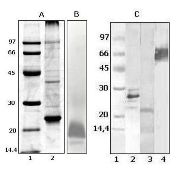
Figure 1.
B. pertussis PL composition analysis; Panel A, SDS-PAGE (polyacrylamide 13 %) followed by R250 Blue Coomasie stain. Lane 1: Low molecular weight markers (97-14.4 kDa), Lane 2: B. pertussis PL. Panel B, SDS-PAGE of B. pertussis PL (polyacrylamide 13 %) followed by a LPS-specific silver stain. Panel C, Western blot of B. pertussis PL using Mabs or serum vs relevant antigens; Lane 1: Low molecular weight markers (97-14.4 kDa), Lane 2: PT anti serum (97/572), Lane 3: FIM 3 Mab (04/156), Lane 4: PRN anti serum (97/558).

