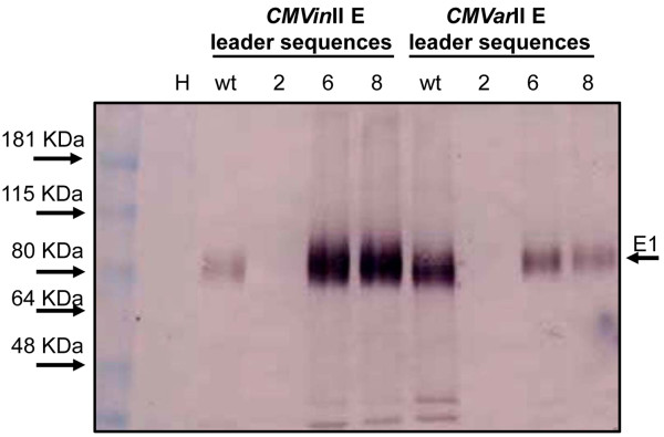
Figure 5.

Comparison of GFP fluorescence from CMVarI G and CMVarII G variants. N. benthamiana leaves were co-infiltrated with A. tumefaciens containing plasmids corresponding to the CMVarI and II G variants (see Table 2) as shown at Panel A. Numbers for leaves in panel at right indicate specific RNA 3 variant. Photographs were taken 6 days post-infiltration under UV light shown as Panel B.
