Figure 9.
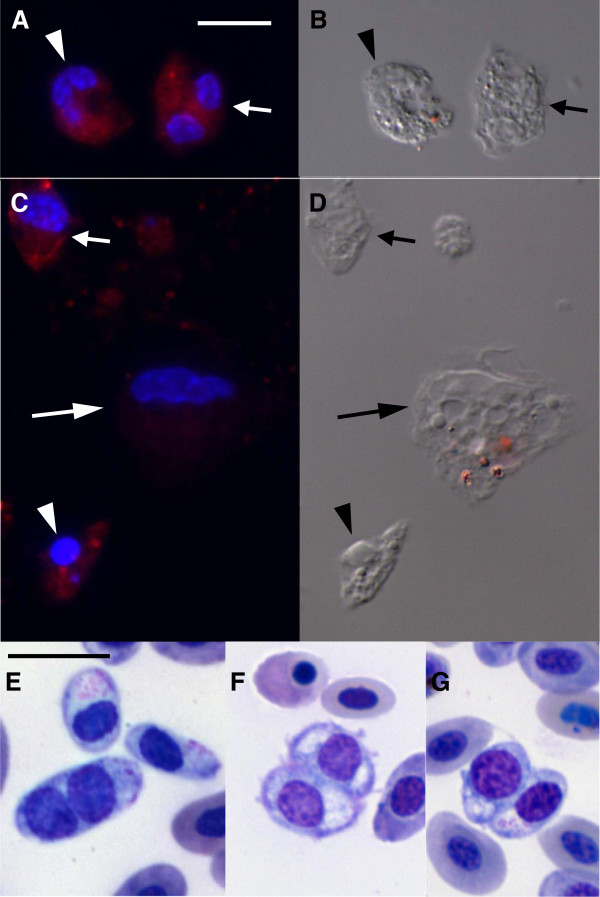
Atypical thrombocytes were positive for the anti-thrombocyte antibody, 11C3, and were rarely detected in stained blood smears. Fluorescence (A, C) and differential interference contrast (B, D) microscopy after labeling cells with the anti-thrombocyte antibody 11C3; red staining is the antibody labeling and blue is DNA. Cells were cultured for 1 (A, B) and 24 h (C, D). Bright field microcopy of stained blood smears (E, F, G). (A, B) Atypical thrombocytes with one nucleus (arrowhead) or two nuclei (arrow) cultured for 1 h were positive for 11C3. (C, D) Normal (arrow) and pyknotic (arrowhead) thrombocytes were positive for 11C3; macrophage (long arrow) was negative. (E- G) Atypical thrombocytes with two nuclei were also observed in blood smears of chickens with high parasitemia. Bar = 10 μm, bar in A and E, is the same for B, C, D and F, G, respectively.

