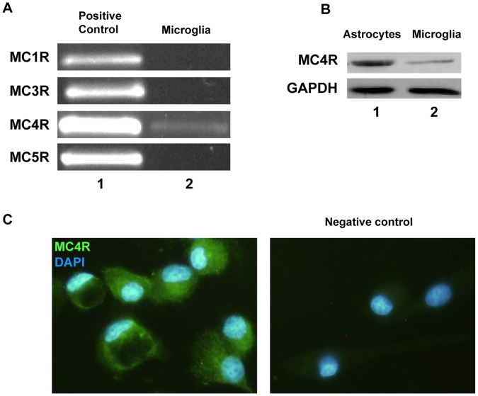
Figure 1. Melanocortin 4 receptor expression in rat microglia.
(A) MCR mRNA expression was assessed by RT-PCR using total mRNA from untreated microglial cells. Genomic DNA was used as a positive control (lane 1). A band close to 595 pb corresponding to MC4R amplification product was detected in primary cultured microglial cells (lane 2, MC4R). We did not detect expression of the other MCR subtypes in these cells (lane 2). (B) MC4R protein expression was evaluated by western blot. Total protein sample from astrocytes was used as a positive control. Results show the presence of a band of nearly 50 KDa in both astrocytes (lane 1) and microglia (lane 2). (C) MC4R was detected in primary microglia by immunocytochemistry. The left panel shows MC4R immunoreactivity in microglial cells. Nuclei were stained with DAPI to identify individual cells. The negative control is shown in the right panel.

