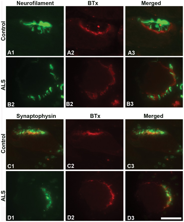
Figure 1. NF-L and synaptophysin at NMJs of EOMs.
Light microscopic images of NMJs from adult controls (A and C) and ALS donors (B and D) double-labeled with α-bungarotoxin (BTx, red) and antibodies against NF-L (green; A1, A3, B1, and B3) or synaptophysin (green; C1, C3, D1 and D3). Note the similar staining patterns between controls and ALS donors. Bar = 20 µm.

