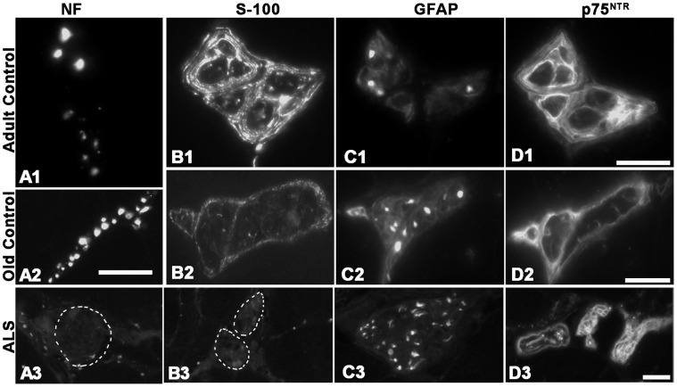
Figure 7. Intramuscular nerve axons in limb muscles.
Light microscopic images of nerve axons from adult (top panel) and elderly (middle panel) controls and from ALS donors (bottom panel) labeled with antibodies against NF-L (A), S100B (B), GFAP (C) and p75NTR (D). Note the absence of NF-L (A3) and S100B (B3) in the nerves (outlined areas) of the ALS donors. Bars = 20 µm.

