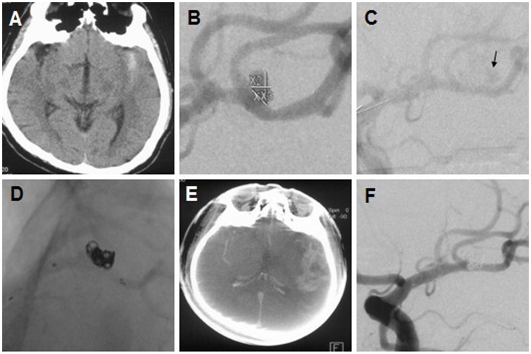
Figure 3. Management of intraprocedural aneurysm rupture.
(A–B) A ruptured MCA-bifurcation aneurysm. (C) Leakage of contrast medium (arrow) and spasmodism of intracerebral arteries. (D) Morphologic presentation of stent and coils. (E) High density haemorrhage area. (F) The 6-month follow-up angiogram showing the total occlusion of the lesion.

