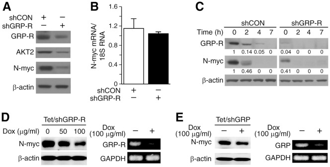
Figure 1. GRP/GRP-R regulated N-myc expression.
(A) N-myc and AKT2 expression in BE(2)-C/shCON and BE(2)-C/shGRP-R cells by Western blotting. (B) MYCN mRNA levels, measured by real-time QRT-PCR, remained relatively unchanged. (C) Cells were serum-starved for 24 h and then re-plated in fresh RPMI media with 10% FBS. Decreased GRP-R expression in shGRP-R cells when compared to shCON cells was confirmed. N-myc expression was also decreased in shGRP-R cells at 0 and 2 h. Protein levels were quantified by densitometric analysis values indicated each band. (D) Inducible GRP-R silencing BE(2)-C/Tet/shGRP-R cells were treated with doxycyclin for 48 h, and then N-myc expression was analyzed by Western blotting. N-myc protein level was correspondingly decreased with GRP-R silencing (left); Decreased GRP-R mRNA was confirmed with RT-PCR (right). (E) Similar to GRP-R silencing, inducible GRP silencing BE(2)-C/Tet/shGRP cells were treated with doxycyclin for 48 h, and then N-myc expression was assessed with Western blotting (left). Inducible knockdown of GRP mRNA was confirmed with RT-PCR (right).

