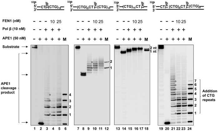
Figure 7. Pol β DNA synthesis during BER of an abasic lesion located at different sites of (CTG)20 repeats.
Pol β DNA synthesis with the substrates containing one or two THF residues located at the 5′-end, or/and in the middle, or at the 3′-end of (CTG)20 repeats was determined in the presence of 10 nM of pol β along with 10 nM and 25 nM FEN1. Lanes 1, 7, 13, and 19 correspond to substrates only. Lanes 2, 8, 14, and 20 correspond to reaction mixtures with 50 nM APE1. Lanes 3−5, 9−11, 15−17, and 21−23 correspond to reaction mixtures with 10 nM pol β in the absence or the presence of 10 nM and 25 nM FEN1. Lanes 6, 12, 18, and 24 correspond to a series of synthesized size markers (M) for illustrating the size of pol β DNA synthesis products. Substrates were 32P-labeled at the 5′-end of their damaged strands. Substrates are illustrated schematically above the gel.

