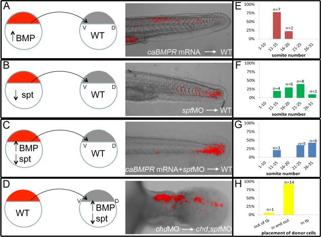
Figure 4. Ability to exit the tailbud is a cell autonomous fate decision.
Cell transplants were performed at 30-50% epiboly stages as diagramed above. Labeled donor cells were placed on the ventral lateral margin of unlabeled host embryos. A-D, Live embryos were photographed using confocal microscopy at 30-48hpf. E-H, Recipient embryos were scored based on the most anterior somite containing donor cells. Somite 1 being the most anterior and 31 being the most posterior. Cells injected with caBMPR mRNA 4ug/mL and placed in a WT host were able to exit the tailbud and differentiate in posterior and anterior tail somites, n=9 (A, E). Cells injected with sptMO 3ug/mL were also able to leave the tailbud in WT background and contribute to tail somites, n=20(B, F). Cells injected with caBMPRmRNA +sptMO were not able to efficiently leave the tailbud in WT backgrounds. A few donor cells could be occasionally found in posterior tail somites and 3 embryos had donor cells in anterior tail somites. However, the majority of transplanted cells were still in the tailbud at 48hpf, n=14 (C, G). When chdMO cells were placed in a chdMO;sptMO background they were able to exit the tailbud and migrate anteriorly, n=15 (D, H).

