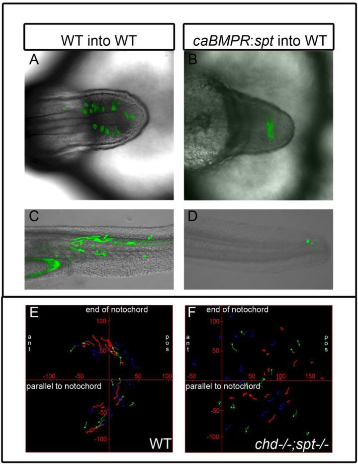
Figure 5. Cell movements are perturbed in cells lacking spadetail and experiencing high BMP levels.
A-B, Top-down view of tailbud at 18hpf with transplanted WT(A) and caBMPR mRNA + sptMO(B) cells in WT background. C-D Lateral view of 48hpf embryos with transplanted WT (C, n=11) and caBMPR mRNA + sptMO (D, n=18) in a WT background. Transplanted WT cells are able to exit the tailbud and contribute to somites (A, C). caBMPR mRNA + sptMO cells remain in the tailbud forming a tight cluster and do not intermix with WT cells or form somites(B,D). E-F, Time lapse images were made of mGFP labeled embryos. Stills were taken every 90 seconds for 1-2 hours of development at 14 somite stage. Manual cell tracking was performed for 10-30 consecutive frames using ImageJ and placed on graphs with X axis parallel to the notochord and Y axis perpendicular to the end of the notochord, tick marks on axis are 50um. Tailbud cells in WT embryos (n=3) display movement away from the midline and anterior migration to form tail somites (E). chd:spt tailbud (n=3) cells do not display a uniform pattern of movement and their migration paths are much shorter (F).

