
Figure 2. KLF5 is required for villus morphogenesis and epithelial differentiation.
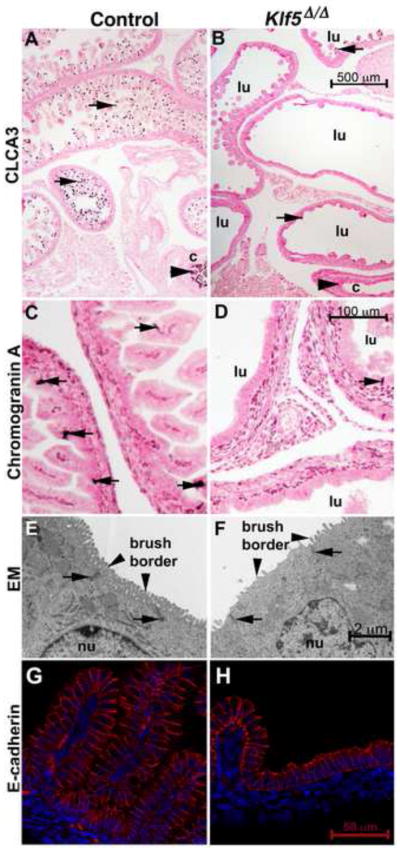
At E18.5, the goblet cell marker CLCA3 stained goblet cells on the villi of the small intestine (arrows in A) and within the colon of control fetuses (arrowhead). Villus-like structures were rarely observed in the Klf5Δ/Δ epithelium (B) and few goblet cells were present in either the intestine (B, arrows) or colon (arrowhead). Chromogranin A positive staining enteroendocrine cells were present in controls (C, arrows) but rare in the Klf5Δ/Δ tissue (D, arrows). The enterocyte brush border was abnormal in the Klf5Δ/Δ epithelium (E, vs. F, arrowheads) as assessed by transmission electron microscopy. The location of tight junctions was similar in both tissues (E, F arrows). E-cadherin staining indicates that like the control (G) the epithelium lining the Klf5Δ/Δ (H) intestine was of columnar morphology on the flattened regions and on the rudimentary villus-like structures. Lumen of intestine (lu), colon (c), nucleus (nu). Scale bars for each stain are present in the Klf5Δ/Δ image.