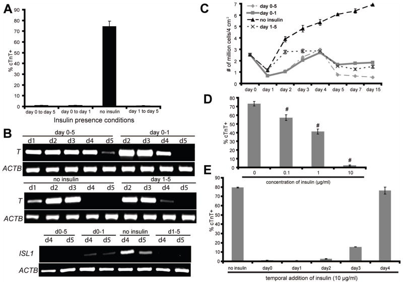
Figure 3.
(A–D) H9 cells were differentiated as described in Fig. 1(A), with insulin present or absent during the indicated stages of differentiation. (A) 15 days after initiation of differentiation, cells were analyzed for cTnT expression by flow cytometry. (B) At different time points, mRNA was collected and RT-PCR analysis of mesendoderm (T) and cardiac gene expression (ISL1) was performed. (C) At different time points, single cells were prepared by Accutase treatment and counted. (D–E) Cardiomyocytes were generated from H9 cells using the protocol described in Fig. 1(A), with RPMI/B27-insulin medium used from day 0 to day 5. (D) At day 1, indicated concentrations of insulin were added into the culture medium and flow cytometry for cTnT expression was performed at day 15. Error bars represent the s.e.m. of eight independent experiments. # p<0.005, each sample with insulin versus without insulin; t test. (E) 10 μg/ml insulin was added to the culture medium at the indicated time points of differentiation. 15 days after initiation of differentiation, cells were analyzed for cTnT expression by flow cytometry.
Error bars represent the s.e.m. of three independent experiments.

