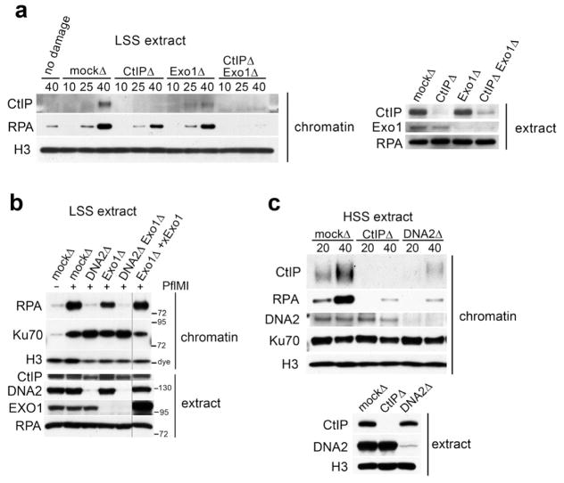
Figure 1.
Contribution of individual resection pathways to resection of chromosomal DNA DSBs. (a) Chromatin was incubated in extract that was depleted of CtIP, Exo1, both, or mock-depleted and isolated at the indicated time (minutes) after addition of 0.05 U/μL PflMI restriction endonuclease, and immunoblotted with the indicated antibodies (“chromatin”, top panels). Aliquots of the depleted extract were immunoblotted with the indicated antibodies to access immunodepletion (“extract”, lower panel). (b) Chromatin was incubated in mock-depleted LSS or LSS immunodepleted of DNA2, Exo1, or DNA2 and Exo1, and chromatin-isolation was performed as in (a). Recombinant Exo1 was added to Exo1-depleted extract as indicated. (c) HSS extract was mock-depleted, or depleted of either CtIP or DNA2, and chromatin-isolation was performed as in (a).

