
Figure 3. MSCs and HUVECs migrate in response to bioactive lipids, and C1P increases adhesion of MSCs.

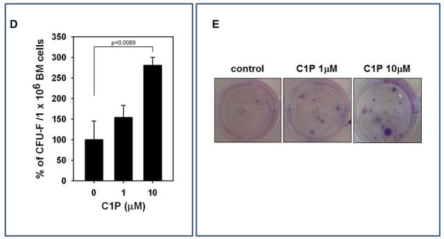
Murine bone marrow-derived multipotent stroma cells (msMSCs, Panel A), HUVECs (Panel B), and human BM-derived multipotent stroma cells (huMSCs, Panel C) were loaded into an upper Transwell chamber (8-μm), with the lower chamber containing different bioactive lipids, such as S1P (0.1 μM), C16-C1P (1 μM), C18-C1P (1 μM), LPA (1 μM), LPC (1 μM) or SDF-1 (low dose (L), 10 ng/ml and high dose (H), 300 ng/ml). Twenty-four hours later, cells remaining in the upper chambers were scraped off with cotton wool, and cells that had transmigrated were stained by HEMA 3 according to the manufacturer’s instructions (Fisher Scientific, Pittsburgh, PA, USA) and counted either on the lower side of the membranes or on the bottom of the Transwell chambers. The data shown represent the combined results from two independent experiments carried out in quadruplicate per group. *p<0.05 Panel D: For the CFU-F assay, whole BM cells were stimulated with C1P and seeded into culture plates. Mouse whole bone marrow cells were stimulated with C18-C1P (1 or 10 μM) for 1 hour and seeded (1 × 10⁶ cells/well, into each of 6 wells). On the next day, non-adherent cells were removed and cultured for 14 days in complete medium. Adherent cells were washed twice with PBS, stained with the HEMA 3 staining kit, and the colonies counted. The data shown represent the combined results from three independent experiments carried out in quadruplicate per group. *p<0.0089 Panel E: Representative stained colonies of adherent cells are shown.