
Figure 6. C1P does not affect proliferation of HUVECs, but triggers capillary-like structure formation in 3D-Matrigel cultures, angiogenesis in vivo in Matrigel implants and enhances adhesion of HUVECs.

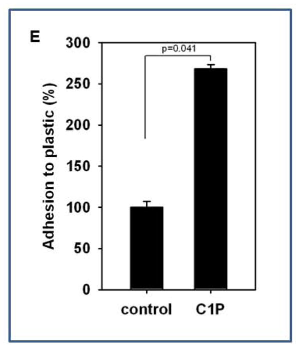
Panel A: HUVECs were cultured with C18-C1P (0.1 or 1 μM) for 1, 2, 3, and 4 days and cells were counted at the indicated time points. The data shown represent the combined results from two independent experiments carried out in quadruplicate per group. Panel B: HUVECs were seeded into Matrigel cultures in the presence of bioactive lipids S1P (0.1 μM), C16-C1P (1 μM), C18-C1P (1 μM), LPA (1 μM), LPC ( 1 μM), SDF-1 (50 ng/ml), or FGF2 (50 ng/ml, positive control) in serum-free media, incubated for 4 hours at 37°C, and the number of tubules counted. *p<0.05 Panel C: Tube formation in the presence of C18-C1P. Panel D: Hemoglobin level in Matrigel implants supplemented with C1P (1 or 10 μM), FGF2 (50 ng/ml, positive control) or in presence of medium alone. Matrigel implants were removed and analyzed 2 weeks later. *p<0.05 Magnification × 100. Panel E: C1P increases HUVEC adhesion. HUVECs were stimulated with C18-C1P (1 μM) for 1 hour and seeded (5 × 10⁴ cells/well, in each of 6 wells); after 3 minutes, non-adherent cells were removed by washing twice with PBS.