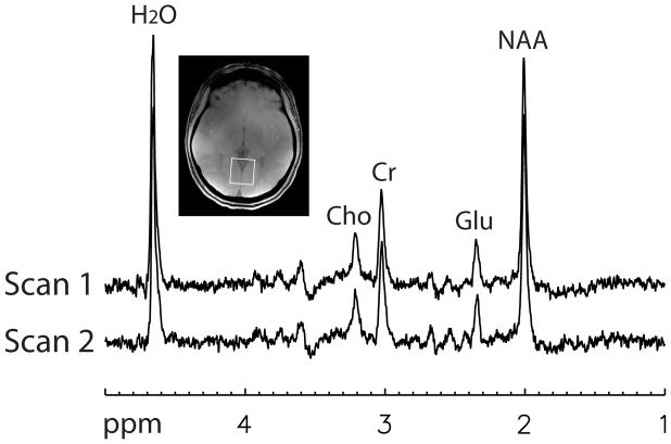
Fig. 3.
Two combined spectra, one for each scan, of the first normal volunteer computed using the GLS method. Each combined spectrum was computed using the 32-channel data from the first acquisition. Voxel size = 3 × 3 × 3 cm3, TR = 2.5 s, TE1 = 37 ms, TE2 = 63 ms, spectral width = 4000 Hz, number of samples = 2048, number of acquisitions = 128.

