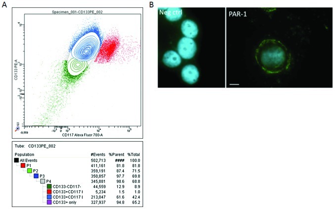
Figure 3.
Immunoanalysis of the ovarian cancer cell line, OVCAR-3. (A) Three subpopulations of OVCAR cells were identified by the cell sorter. They were CD133+ (65.2%), CD133+/CD117+ (42.4%) and CD133− (8.9%). (B) PAR-1 immunocytochemistry. OVCAR cells were grown on glass bottom chamber slides, fixed and successively incubated with PAR-1 antibody, appropriate biotinylated secondary antibody and FITC-streptavidin. Isotype antidodies were used in parallel and the nuclei were DAPI-labeled. Initial magnification, ×400. Scale bar represents 10 μm.

